BioengineeredTumor

Biomimetic tumor engineering
to enhance drug discovery

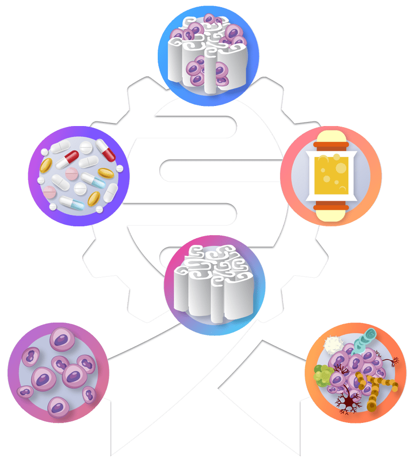
Stepping up cancer research and drug development with our tumor engineering approach, based on 3D cancer cell cultures supported by biomimetic biomaterials and bioreactors.

Supported by:

The Science Fund of the Republic of Serbia, established in March 2019, supports science and research by providing funding and fostering an environment for their continuous development. It aims to advance scientific activities in Serbia, essential for progressing toward a knowledge-based society.

What is BioengineeredTumor?

About

The BioengineeredTumor project aims to upgrade the cancer research and drug testing by development of a sufficiently simple, but relevant, adaptable and scalable platform suited to the use by scientists without technical expertise for in vitro studies of cancer cells. Envisioned applications of such a platform are in: (I) anti-cancer drug discovery and validation, (II) development of personalized medical treatments, and (III) cancer research. Our multidisciplinary team composed of engineers, molecular biologists, medical doctors and pharmacists is well suited to address the complex problem of recapitulating tumor features in vitro by bringing in both engineering and cell biology aspects in cancer cell cultures.

Collaborating Institutions:

University of Belgrade
Faculty of Technolgy and Metallurgy

Innovation Center of the Faculty of Technology and Metallurgy

Institute of molecular genetics and genetic engineering

Institute for Biological Research “Siniša Stanković”

University of Belgrade
Faculty of  Pharmacy

University of Belgrade
Faculty of  Medicine

Meet The Team

Our Team

Behind The Project

Meet the individuals behind the BioengineeredTumor project. Our diverse team of engineers and life scientists from various institutions passionately pursues innovation in cancer research. 

Contact Us:

Thank you for your interest in our project! For more updates and direct communication, feel free to connect with us on our social media platforms or send us an email. We appreciate your support in our endeavor to advance cancer research. Together with our collaborating institutions and our main sponsor, the Science Fund, we’re making strides towards a brighter future in healthcare. We look forward to hearing from you.

Malignant Tumor Cells

Several human and animal cell lines will be used in the project. For the carcinoma model, cervical carcinoma cell lines (SiHa and HeLa) and human lung carcinoma NCI-H460 will be used. For the osteosarcoma model, the human cell line Saos-2 and the mouse cell line K7M2-vt will be employed. By selecting these cell lines, the aim is to determine the effects of 3D cultures depending on the type of cancer cells (carcinomas, osteosarcomas) and the origin of the cells (human, animal).

Biomaterials

Cells will be cultured within biomimetic scaffolds designed to mimic the extracellular matrix of tumors. Specifically, alginate hydrogels will be produced in the form of microfibers (diameter < 1 mm) to immobilize carcinoma cells, providing a soft and hydrophilic environment. Conversely, macroporous composite alginate carriers with a mineral phase will be used to immobilize osteosarcoma cells, thereby providing the composition and structure relevant to bone tissue. Hydroxyapatite (HAp) and bioactive glass (BAG) particles will be used as the mineral phase to mimic the environment in mature bone tissue and remodeling bone tissue, respectively. In the presence of biological fluids, as well as in cell culture medium, BAG transforms into HAp. Microfibers will be produced by simple manual extrusion of the cell suspension in Na-alginate into a gelling solution containing Ca or Ba ions, which replace Na ions and thus ensure alginate gelation and cell immobilization within the gel. Macroporous composite alginate carriers with a mineral phase in the form of particles will be prepared by controlled gelling of an alginate and powder mixture, followed by lyophilization of the obtained hydrogel, and then partial rehydration in cell culture medium. Afterward, cells will be manually seeded onto the carriers, and the procedure will be optimized in terms of seeding efficiency, cell distribution within the carriers, and cell viability retention. All carriers will be characterized in terms of composition and structure using standard techniques (microscopy and spectroscopy methods).

Biomimetic Bioreactors

The disposable perfusion bioreactor (“3D Perfuse”, Innovation Center of the Faculty of Technology and Metallurgy, Belgrade), developed by the team at the Faculty of Technology and Metallurgy, is the third component of the 3D tumor model. The bioreactor provides direct flow of the medium through the sample placed in the bioreactor chamber, which improves mass transfer to the cells within the carrier.

3D Cell Cultures

3D cancer cell cultures based on the use of biomimetic carriers and a perfusion bioreactor will be characterized and optimized in terms of physiological aspects of cells such as morphology, viability, proliferation, and immunological and genetic expression. The goal of these studies is to demonstrate differences between various types of cancer cells, cells cultured in 2D and 3D environments, and cells derived from animals and humans. The cultures will be evaluated in terms of: cell viability and proliferation, metabolic activity, apoptosis, cell distribution within carriers, and morphological features, cytokine profiles in the cell culture medium, as well as differentially expressed genes. Mathematical modeling will be applied to define mass transfer rates and shear stress in flow cultures. The outcome of this phase will be optimized 3D models that can support carcinoma and osteosarcoma cell cultures over an extended period (up to 28 days) while retaining key characteristics of cancer cells.

Anti-Cancer Drug Testing

Optimized 3D models for carcinoma and osteosarcoma cell cultures will be evaluated in studies of the effects of antitumor drugs. In the initial phase, short-term cultures (up to 7 days) will be used as relevant for the rapid preclinical pharmacological evaluation of new drug candidates, while long-term cultures (up to 28 days) will be used to mimic clinically relevant drug administration regimes. In these studies, cisplatin, doxorubicin, 5-fluorouracil, and/or paclitaxel will be used as standard drugs commonly employed in clinical practice for treating various types of cancer including carcinomas and osteosarcoma. A critical analysis of the results obtained, compared to literature data on the effects of standard antitumor drugs, will clearly demonstrate the relevance of the developed 3D tumor models for application in preclinical pharmacological studies.

Standardized 3D Tumor Model

The BioengineeredTumor project aims to establish a controlled platform for 3D cancer cell cultures and antitumor drug testing that will be adaptable to other types of cells, as well as autologous cells and tissues from patients. By using a systematic and integrated methodology to comprehensively define key model components in combination with the development/adaptation of analytical methods for reliable characterization of cells within 3D carriers, the goal is to comprehensively characterize biomaterials in terms of geometry and structure, as well as hydrodynamic conditions in the flow bioreactor to link cultivation condition parameters with all key characteristics of the cultured cancer cells. Thus, the conceived 3D platform for cancer cell cultures is ultimately intended for use as i) a preclinical pharmacological tool for testing antitumor drugs and ii) a future medical device for identifying the most effective personalized cancer treatment protocols.